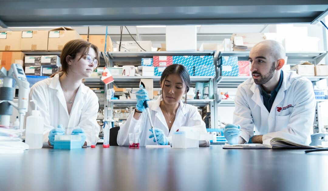
For researchers studying heart disease, lab-grown heart cells are an essential tool. Created from human stem cells, they offer a powerful way to model disease, test therapies, and better understand heart function. However, the challenge remains that these cells behave...
For children living with cardiomyopathy, heart failure is not always caused by the heart losing its ability to pump. In many cases, the problem is more subtle, but just as serious: the heart becomes stiff and struggles to relax and fill properly between beats. This...
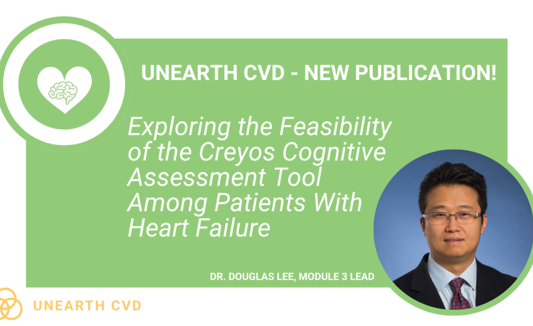
New findings from the UNEARTH CVD research team have shed light on an important but often overlooked aspect of cardiovascular care: the cognitive health of people living with heart failure. Published in Cureus, this new publication is the first study to examine...
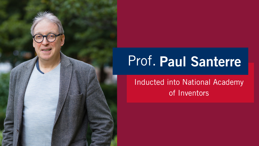
The Ted Rogers Centre for Heart Research is proud to celebrate an extraordinary milestone in the career of Dr. Paul Santerre, who has been elected as a Fellow of the National Academy of Inventors (NAI), the highest honour bestowed upon academic inventors. This...
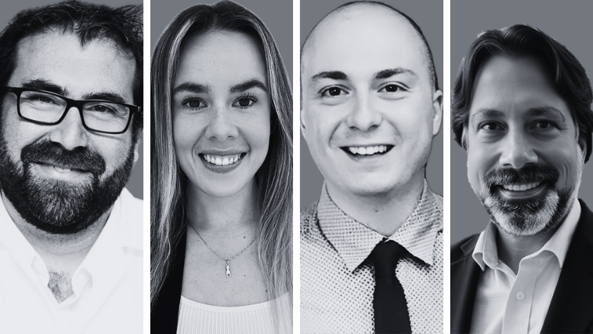
Entrepreneurship for Cardiovascular Health Opportunities (ECHO) is a 12-month national training program supporting cardiovascular research commercialization through education, mentorship, networking, and funding. Led by a diverse team of experts, ECHO fosters...
Entrepreneurship for Cardiovascular Health Opportunities (ECHO) is a 12-month national training program supporting cardiovascular research commercialization through education, mentorship, networking, and funding. Led by a diverse team of experts, ECHO fosters...
3D models of heart failure… stiffened hearts in diabetes…. saving infant lives
After a heart attack, this peptide protects the heart from further injury
Featured Event
2026 Heart Failure Symposium
Virtual Library
Visit our YouTube channel filled with world-class heart failure educational sessions on diverse topics
Investigating Heart Failure
Personalized Medicine
Equitable Access to Care
Translating Innovation
For Patients
For Clinicians
For Researchers
Exploring the Heart’s Hidden Layers: Advances in Cardiac Fibroblast Diversity
In the realm of cardiac health, an exciting new study has cast light on the intricate details of our hearts, revealing significant differences between the left and right ventricles’ fibroblasts. This research, conducted by a team of dedicated scientists from the University of Toronto and the Ted Rogers Centre for Heart Research’s Translational Biology Program (TBEP), has opened new doors in understanding how our hearts respond to injury, potentially paving the way for tailored treatments for heart failure.
Cardiac fibrosis, a condition characterized by the thickening and stiffening of the heart tissue, plays a pivotal role in heart failure. Cardiac fibroblasts (CFs), the star players in this process, have traditionally been viewed as a uniform group. However, this study, building upon existing work in this area, has now unveiled their surprising diversity, challenging long-held beliefs in the field.
Using state-of-the-art RNA sequencing techniques on mice models, the team meticulously analyzed cardiac fibroblasts from both the left and right ventricles of the heart. Mice have hearts with anatomical similarities to those of humans, making them valuable models for cardiac research by replicating the structures present in human hearts. Their findings revealed distinct transcriptomic signatures, highlighting a complex landscape of eight fibroblast subpopulations, including two that had not been previously discovered. Specifically, the results showed 422 differentially expressed genes between left ventricle- and right ventricle-derived CFs, which is new information for the field coming out of this study. These differences are not just academic; they have profound implications for how we understand and treat heart diseases.
Among the most striking discoveries was that fibroblasts from each ventricle appear to be primed differently for activation following injury. In the left ventricle, there are greater numbers of pro-fibrotic fibroblasts following injury, accelerating the process of fibrosis, which is the wound healing response where excessive connective tissue can lead to scarring and a loss of function. The left ventricle is typically impacted during cardiac arrests, or heart attacks. In contrast, the right ventricle, typically implicated in cumulative pressure overload in the heart due to congenital defects, develops fewer of these pro-fibrotic fibroblasts, which may lead to a more moderated fibrotic response.
This research also sheds light on the potential for sex-specific differences in cardiac fibrosis, suggesting that men and women could respond differently to heart injuries. As all experiments were performed in both male and female mice, the results suggest that sex influences the number of injury-induced fibroblasts present, both before and after injury. The injury-induced fibroblasts are best known for producing high levels of stiff extracellular matrix proteins, which may explain the sex differences in both mouse and human scar development. Such insights are invaluable for developing personalized medicine, where treatments can be tailored not just to the specific ventricle affected but also to the patient’s sex.
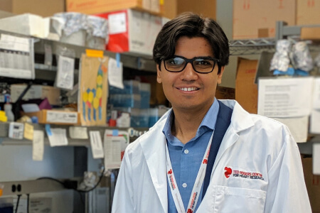
PhD candidate Michael Dewar, lead author of the study.
The implications of this study are far-reaching, according to the publication’s lead author, PhD candidate Michael Dewar, of Prof. Scott Heximer’s lab. “By uncovering the hidden diversity of cardiac fibroblasts, researchers can now explore new therapeutic targets for treating heart failure. This knowledge brings us one step closer to interventions that could halt or even reverse the progression of heart diseases, and ultimately benefit millions of people worldwide.”
Thanks to funding from the Ted Rogers Centre for Heart Research and TBEP, Mr. Dewar and the Heximer team were able to access both cutting-edge technology and experts in fields such as echocardiography, a diagnostic test that uses ultrasound waves to create images of the heart, allowing the team to learn more about fibroblasts in a shorter period of time. As a result, this work may be able to inform key time windows for responding to different cardiac issues knowing how the fibroblasts react differently based on injury.
As we continue to unravel the mysteries of the heart, studies like this underscore the importance of looking beyond the surface and questioning long-held beliefs, venturing into the cellular level to understand the full complexity of our bodies. The future of cardiac care is bright with scientists like Mr. Dewar, who are leading research that is paving the way for innovations that could transform how we treat heart diseases, making personalized, effective treatment not just a possibility but a reality.