For researchers studying heart disease, lab-grown heart cells are an essential tool. Created from human stem cells, they offer a powerful way to model disease, test therapies, and better understand heart function. However, the challenge remains that these cells behave...
For children living with cardiomyopathy, heart failure is not always caused by the heart losing its ability to pump. In many cases, the problem is more subtle, but just as serious: the heart becomes stiff and struggles to relax and fill properly between beats. This...
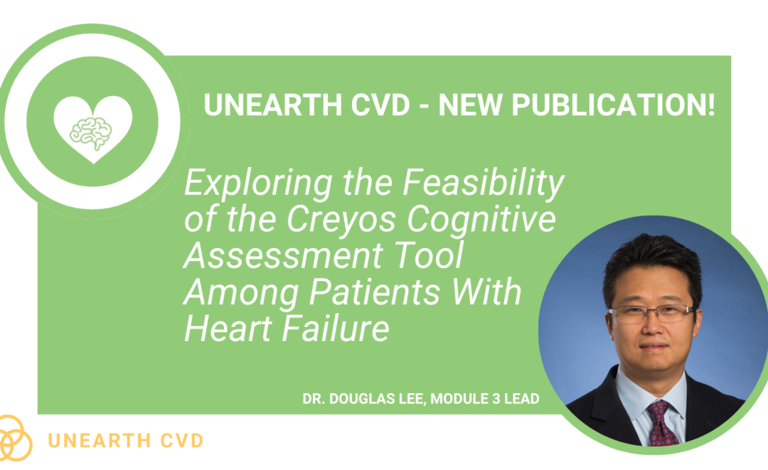
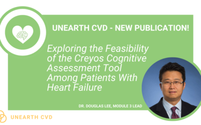
New findings from the UNEARTH CVD research team have shed light on an important but often overlooked aspect of cardiovascular care: the cognitive health of people living with heart failure. Published in Cureus, this new publication is the first study to examine...
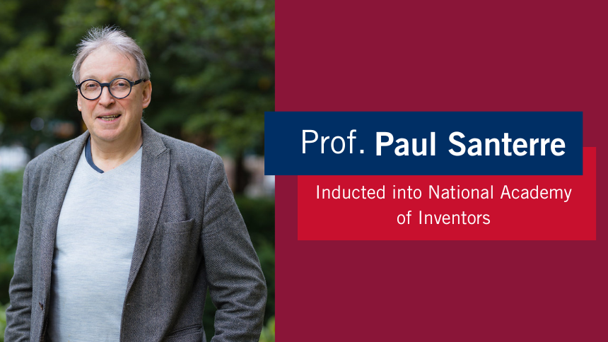
The Ted Rogers Centre for Heart Research is proud to celebrate an extraordinary milestone in the career of Dr. Paul Santerre, who has been elected as a Fellow of the National Academy of Inventors (NAI), the highest honour bestowed upon academic inventors. This...
Entrepreneurship for Cardiovascular Health Opportunities (ECHO) is a 12-month national training program supporting cardiovascular research commercialization through education, mentorship, networking, and funding. Led by a diverse team of experts, ECHO fosters...
Entrepreneurship for Cardiovascular Health Opportunities (ECHO) is a 12-month national training program supporting cardiovascular research commercialization through education, mentorship, networking, and funding. Led by a diverse team of experts, ECHO fosters...
3D models of heart failure… stiffened hearts in diabetes…. saving infant lives
After a heart attack, this peptide protects the heart from further injury
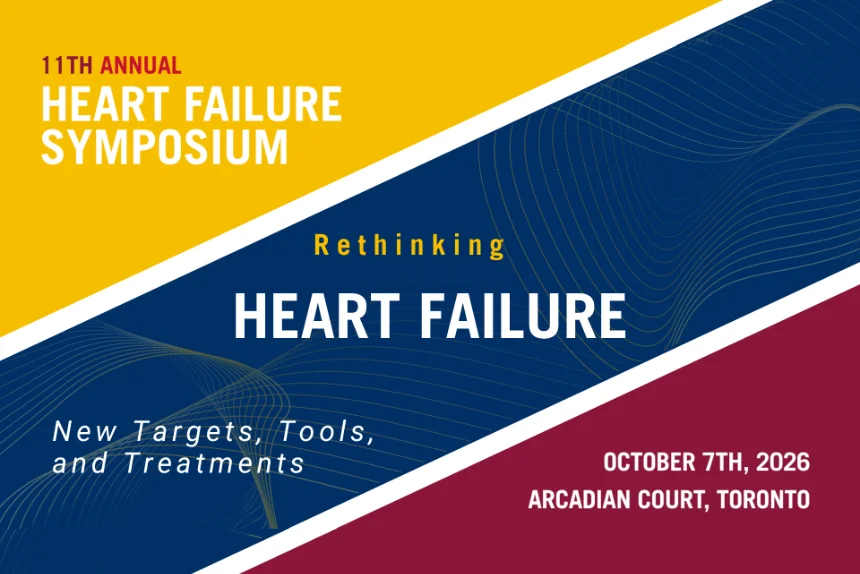
Featured Event
2026 Heart Failure Symposium
Virtual Library
Visit our YouTube channel filled with world-class heart failure educational sessions on diverse topics
Investigating Heart Failure
Personalized Medicine
Equitable Access to Care
Translating Innovation
For Patients
For Clinicians
For Researchers
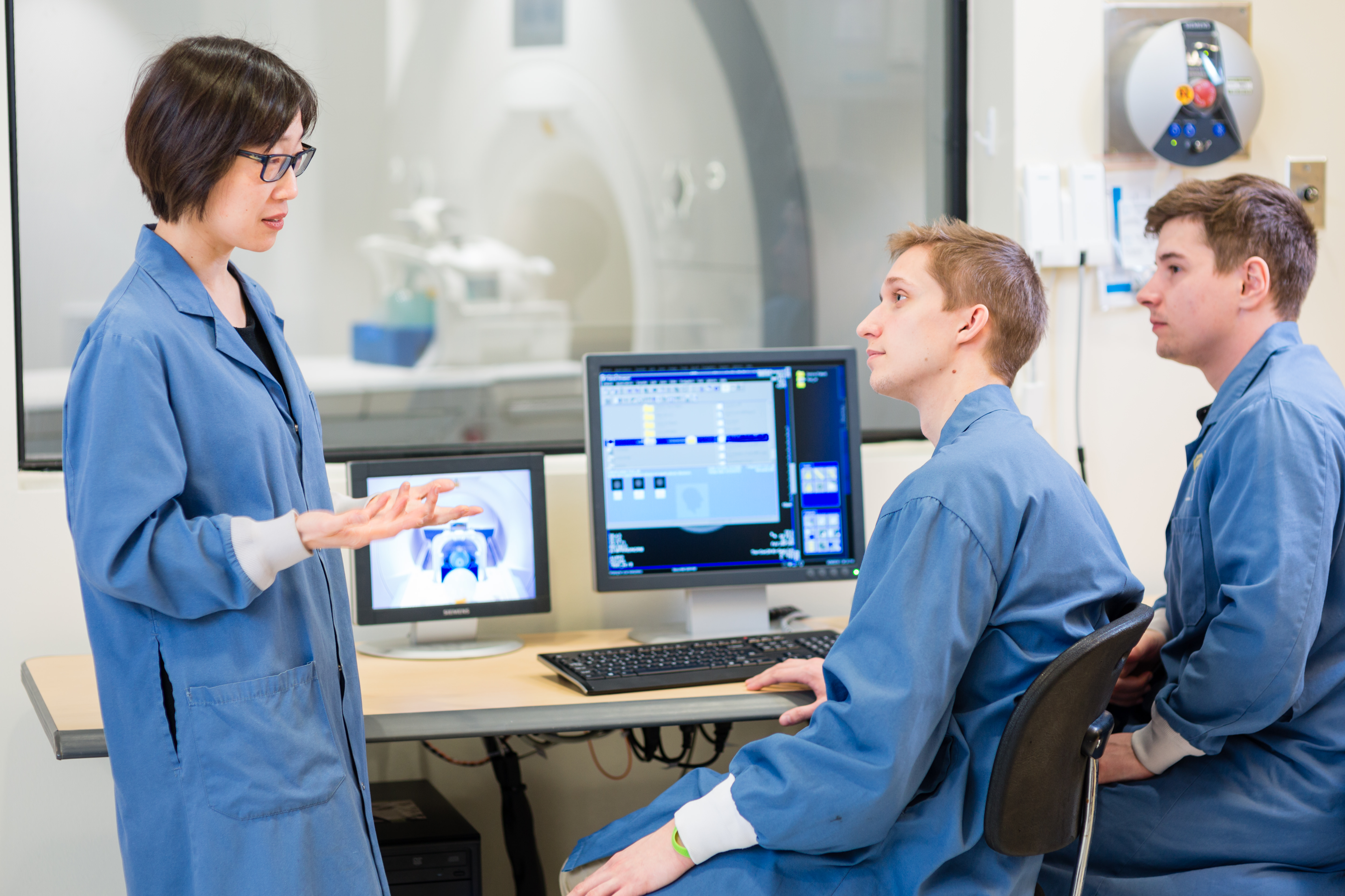
Advanced MRI for CVD diagnosis and cardiac regeneration
Magnetic resonance (MR) imaging is a diagnostic modality that most of us are familiar with. I have known many people, outside of my workplace, who themselves had an MR exam or had a friend or relative undergo one to check out a sports injury, to diagnose cancer, or to follow up after a stroke. Regardless of the reason for which an MR exam is prescribed, most people still think of it as an anatomical imaging modality – where certainly it excels compared to other diagnostic approaches.
(Right click and select “Loop” for above video)
Yet MR can do so much more. It can image tissue function, such as identifying ischemic tissue and measuring microvascular perfusion. It can capture the mechanical motion of the heart. It can visualize therapeutic cells that are introduced into the body. It has the potential to monitor many ingredients that are being investigated for tissue engineering and regenerating new heart tissue. The advantage of MR imaging? It can do all this non-invasively, without ionizing radiation, and in one exam setting.
Toward growing new heart tissue
Our lab in the Translational Biology & Engineering Program of the Ted Rogers Centre for Heart Research is focused on developing new MR technologies for improved cardiac diagnostics and taking these capabilities to investigate novel therapies to regenerate new, healthy heart tissue.
We are devising new contrast mechanisms to enable long-term MR cell tracking in vivo, so that we can truly determine the fate and function of stem cells delivered to the infarct zone. We will soon be translating to cardiac imaging a technology we have developed for looking at microvessel functional dynamics, for which a non-invasive alternative currently does not exist.
This technology will provide important insight into the health of the microvascular bed at early timepoints before an adverse event occurs, and will provide functional assessment of the efficacy of different regeneration strategies. We have also demonstrated new methods by which we can monitor biomaterials used to support tissue regeneration and are aiming to expand the repertoire of materials we can monitor non-invasively. Advanced computing capabilities are being built in our lab to enable the computation of functional and physiological “maps” in a matter of seconds, an important requirement for clinical translation.
Taken together, these technologies are interwoven to build an enabling platform for several regeneration strategies we are currently investigating for growing new heart tissue.